


The Eye
A sense organ that reacts to light and allows vision
Parts of the Eye
The eye is made up of three coats. The outermost coat consists of the cornea and the sclera; the middle vascular layer contains the main blood supply to the eye and consists of the choroid, the ciliary body, and the iris. The innermost layer is the retina.
Cornea: The transparent front part of the eye that covers the iris, pupil, and anterior chamber. The cornea with the anterior chamber and lens refracts light with the cornea. This accounts for approximately two-thirds of the eye’s total optical power.
Sclera: is the opaque, fibrous, protective outer layer.
Choroid: is the vascular layer of the eye that lies between the retina and the sclera. This layer of tissue is made up almost entirely of blood vessels. These blood vessels supply oxygen and nutrients to the outer part of the retina.
Ciliary body: A part of the middle layer of the wall of the eye. The ciliary body includes the ring-shaped muscle that changes the size of the pupil and the shape of the lens when the eye focuses. It also makes the fluid that fills the eye
Iris: controls the amount of light that enters the eye by opening and closing the pupil
Retina: a light-sensitive layer of tissue lining the surface of the eye. It captures light sent through the cornea and crystalline lens. It then creates an image by triggering nerve impulses that pass to various visual centers of the brain via the optic nerve.
Within the globe or interior portion of the eye, there are three spaces: the anterior chamber, posterior chamber, and the vitreous chamber.
Anterior Chamber: The space in the eye that is behind the cornea and in front of the iris.
Posterior Chamber: The space in the eye behind the iris and in front of the lens. The posterior chamber is filled with a watery fluid known as the aqueous humor, or aqueous
Vitreous Chamber: The vitreous chamber is the largest of the three chambers and is located behind the lens and in front of the optic nerve. This chamber is filled with a thick, clear gel-like substance called the vitreous humor
Aqueous Humor: the clear fluid filling the space in the front of the eyeball between the lens and the cornea
Vitreous Humor: The vitreous body is the clear gel that fills the space between the lens and the retina of the eyeball of humans and other vertebrates. It is often referred to as the vitreous humor or simply "the vitreous.
Limbus: the border or margin of a structure, especially the junction of the cornea and sclera in the eye.
Uvea: the pigmented layer of the eye, lying beneath the sclera and cornea, and comprising the iris, choroid, and ciliary body.
Optic Nerve: The optic nerve transmits all visual information including brightness perception, color perception and contrast. The job of the optic nerve is to transfer visual information from the retina to the vision centers of the brain via electrical impulses.
Crystalline lens: a transparent and biconvex structure. Along with the cornea, it helps to refract light to focus on the retina. By changing shape, the lens functions to change the focal distance of the eye. This happens so that it can focus on objects at various distances.
The macula and fovea are small areas within the retina that contain the rods and cones. These structures determine the color and shape of the image you are viewing.
A layer at the back of the eyeball containing cells that are sensitive to light and that trigger nerve impulses that pass via the optic nerve to the brain, where a visual image is formed. Its job is to receive light from the lens, convert it to neural signals and transmit them to the brain for visual recognition.
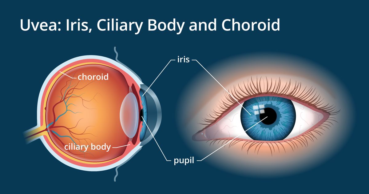
The uvea is the pigmented middle layer of the eyeball. It has three segments:
Iris: In addition to giving the eye its color, the iris acts like the diaphragm of a camera and controls the size of the pupil.
ciliary body: holds the lens of the eye in place. It is connected to the lens with a network of many tiny ligaments that suspend the lens in place behind the pupil.
the choroid: The posterior portion of the uvea that contains many tiny blood vessels and has the vital role of nourishing the retina.
The crystalline lens of the eye is a natural lens which produces one third of the eye's total optical power and focuses light into an image on the retina (the light-sensitive tissue at the back of the eye)
Anatomic Directions
Anatomic Planes
Anatomical Orientation and Directions
Human Anatomy and Physiology Lab (BSB 141) by lumen Learning
Opthalmic Instrumentation
Keratometer: also known as an ophthalmometer, is a diagnostic instrument for measuring the curvature of the anterior surface of the cornea, particularly for assessing the extent and axis of astigmatism.
Automated Corneal Topographer: also referred to as Photokeratoscopy and Videokeratoscopy, is a technique that is used to map the curved surface of the Cornea. This can help measure the quality of vision as well as assist in LASIK surgery and the fitting of contact lenses.
Anatomy, Physiology & Pathology of the Human Eye
Provides images and description of the anatomy, physiology, and pathology of the human eye.
Anatomy of the Human Body by Henry Gray
Features 1,247 vibrant engravings—many in color—from the classic 1918 publication, as well as a subject index with 13,000 entries ranging from the Antrum of Highmore to the Zonule of Zinn.
BioDigital
Hailed as the equivalent of Google Maps for the human body, the BioDigital Human is a scientifically accurate cloud based virtual body that empowers everyone to learn about health and medicine in an entirely new visual format.
Anatomy and Physiology
Anatomy and Physiology is a dynamic textbook for the two-semester human anatomy and physiology course for life science and allied health majors. The book is organized by body system and covers standard scope and sequence requirements.
AnatomyZone
Free anatomy videos, questions, flashcards, tutorials, and a 3D atlas.
Virtual Anatomy and Physiology Classroom
Lectures, PPT presentations, and more
All about Eyes
Understand The Different Parts Of Your Eye
Human Eye Anatomy
Article in encyclopedia Britannica online written by Hugh Davson
Crystalline Lens and Cataract by Joah F. Aliancy, MD and Nick Mamalis, MD
Online article
Anatomy, Physiology and Pathology of the Human Eye
This site includes descriptions, functions, and problems of the major structures of the human eye: conjunctiva, cornea, iris, lens, macula, retina, optic nerve, vitreous, and extraocular muscles. A glossary is included. There also is a test for color deficiency and two short quizzes.


